

Schedule Appointment


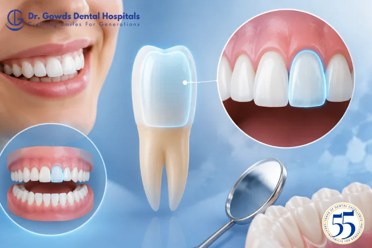
When dentists describe a specific area of a tooth, they often use anatomical terms that may be unfamiliar to patients. One of these terms is labial tooth surface.
The labial tooth surface is the part of a tooth that faces the lips. The term is most commonly used when describing the outer surface of the front teeth, particularly the incisors and canines.
Understanding this terminology can make dental records, treatment explanations, and discussions about tooth anatomy easier to follow.
The labial surface is also relevant to several dental concerns, including plaque accumulation, staining, tooth decay, enamel changes, dental trauma, orthodontic treatment, and cosmetic dentistry.
The labial tooth surface is the surface of a tooth that faces toward the lips.
The word “labial” is derived from the Latin word labium, meaning lip.
In simple terms:
Labial = toward the lips.
For example, the front-facing surface of an upper central incisor that faces the upper lip is its labial surface.
The term is particularly useful when describing the front teeth because their outer surfaces are oriented toward the lips.
The term labial is primarily used when describing the outer surfaces of the anterior teeth.
These include:
For posterior teeth, such as premolars and molars, dentists generally use the term buccal surface because these teeth face the cheeks.
This distinction helps dentists describe the exact location of a cavity, filling, fracture, stain, restoration, or other dental finding.
Labial and buccal are both terms used for tooth surfaces that face outward, but they refer to different anatomical areas.
The surface that faces the lips.
It is primarily used when describing the front teeth.
The surface that faces the cheek.
It is commonly used when describing premolars and molars.
“Facial” is a broader term that refers to the tooth surface facing the face. Depending on the tooth, this can refer to either the labial or buccal surface.
Therefore, the labial surface can be considered a type of facial surface.
The labial and lingual surfaces are located on opposite sides of a front tooth.
Labial surface: faces the lips.
Lingual surface: faces the tongue.
For example, on an upper front tooth, the surface visible when you smile is generally the labial surface, while the surface facing the tongue is the lingual surface.
This distinction allows dentists to describe the precise location of a dental finding.
The labial surface is particularly important for the appearance of the front teeth because it is visible during smiling and speaking.
It can also be involved in several common dental concerns.
Because the labial surface is highly visible, discoloration in this area can be noticed easily.
Tooth discoloration may be associated with:
Not every discoloration is an external stain. Some color changes originate within the tooth and may require professional evaluation.
Plaque can accumulate on the labial surface just as it can on other areas of the teeth.
If plaque is not removed regularly, it can contribute to tooth decay and gum inflammation.
The area close to the gumline deserves particular attention because plaque can accumulate where the tooth and gum meet.
Cavities can develop on the labial surface, although the risk varies between individuals.
Factors that can influence the risk of tooth decay include:
Early tooth decay may not cause pain. A white, brown, or dark area on the front of a tooth can also have causes other than decay, so unexplained changes should be evaluated by a dentist.
The smooth outer surface of a front tooth may appear easy to clean, but plaque can still accumulate along the gumline and in areas that are not effectively reached during brushing.
A consistent oral hygiene routine can help maintain the health of the labial surfaces.
This includes:
The goal is effective and consistent plaque removal rather than aggressive scrubbing.
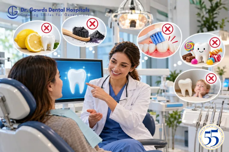
Tooth enamel is strong, but brushing with excessive force can contribute to tooth wear, particularly when combined with abrasive products.
Aggressive brushing can also contribute to gum recession.
When the gums recede, part of the tooth root may become exposed. Unlike the crown, the root surface is not covered by enamel, which can make it more susceptible to sensitivity and wear.
Using a soft-bristled toothbrush and a gentle brushing technique can help reduce unnecessary stress on the teeth and gums.
The labial surface of a front tooth meets the surrounding gum tissue.
When plaque accumulates along the gumline, the gums can become inflamed.
Possible signs of gum inflammation include:
Persistent or recurring symptoms should not simply be considered normal. A dental examination can help identify the underlying cause.
Because the labial surfaces of the front teeth are highly visible, they are commonly involved in cosmetic dental procedures.
Depending on the condition of the teeth and the patient’s goals, treatment may include:
Professional or dentist-guided whitening may help improve certain types of external tooth discoloration.
Veneers are thin restorations placed on the front surfaces of teeth. In selected cases, they can improve the appearance of tooth color, shape, or minor aesthetic irregularities.
Tooth-colored composite material may be used to repair small chips, reshape teeth, or improve the appearance of selected areas.
A crown covers a tooth more extensively and may be recommended when a tooth is significantly damaged or structurally weakened.
The appropriate treatment depends on the underlying dental condition, not appearance alone.
The labial surfaces of teeth are also relevant in orthodontic treatment.
Traditional fixed braces are commonly attached to the outer surfaces of teeth, meaning the brackets can be visible when a person smiles.
During orthodontic treatment, maintaining good oral hygiene around the brackets is particularly important because these areas can be more difficult to clean.
Poor plaque control during orthodontic treatment can increase the risk of enamel demineralization, including white spot lesions, and tooth decay.
White spots on the front surface of a tooth can have several possible causes.
These may include:
A white mark does not automatically mean that the tooth has a cavity.
A dentist may consider the appearance, location, texture, dental history, and other clinical findings when determining the likely cause.
This is particularly important when white spots develop during or after orthodontic treatment.
Front teeth can be injured during sports activities, falls, accidents, or direct blows to the face.
Because the front teeth are exposed toward the lips, injuries affecting them may be particularly noticeable.
Dental trauma can result in:
Even when a tooth appears to have only a small chip, an examination may be appropriate after significant trauma because damage can extend beyond what is immediately visible.
Dentists use tooth-surface terminology to describe findings accurately.
A dental record may refer to a restoration, discoloration, fracture, lesion, or other finding on the labial surface.
This terminology helps distinguish the affected area from other tooth surfaces, including:
Understanding these terms can make dental records and treatment explanations easier for patients to follow.
Protecting the labial surfaces does not require a complicated routine.
Brush twice daily with fluoride toothpaste and a soft-bristled toothbrush.
Applying more force does not necessarily clean teeth better. Gentle brushing can help protect the enamel and gums.
A toothbrush cannot effectively clean every area between the teeth. Use an appropriate interdental cleaning method as part of your daily routine.
Frequent consumption of sugary foods and drinks can increase the risk of tooth decay.
A properly fitted mouthguard can help reduce the risk of dental injuries during sports and activities where facial impacts are possible.
If a new white, brown, or dark area appears on a tooth, professional evaluation can help determine whether it is staining, an enamel change, decay, or another condition.
Routine dental examinations can help identify early changes and allow appropriate preventive or restorative care when needed.
A change on the labial surface does not always indicate a serious dental problem, but certain signs deserve professional evaluation.
Consider scheduling a dental examination if you notice:
Early assessment can help distinguish between a cosmetic concern and a condition that requires treatment.
At Dr Gowds Dental Hospitals, identifying the exact location and cause of a dental concern is an important part of treatment planning.
Whether a visible change affects the labial surface, another tooth surface, or the surrounding gum tissue, a dental examination can help determine the underlying cause.
Depending on the diagnosis, treatment may involve preventive care, restorative dentistry, cosmetic procedures, orthodontic treatment, or other appropriate dental services.
Rather than treating a visible surface change based only on appearance, professional evaluation can help determine what is happening in and around the affected tooth.
Visit us at-
Dr gowds dental hospital Nanakramguda
Dr gowds dental hospital Gachibowli
Dr gowds dental hospital madhapur
The labial tooth surface is the part of a tooth that faces the lips. It is particularly relevant to the front teeth, where this surface is visible during smiling and speaking.
Staining, plaque accumulation, tooth decay, enamel changes, dental trauma, orthodontic brackets, and cosmetic dental procedures can all involve the labial surface.
Understanding the difference between labial, buccal, lingual, mesial, and distal surfaces can also make dental terminology easier to understand.
If you notice a new change on the front surface of a tooth, do not automatically assume it is only a cosmetic issue. A dentist can determine whether the change is related to staining, enamel, decay, trauma, gum problems, or another condition and recommend appropriate care.
The labial tooth surface is the part of a tooth that faces the lips. The term is primarily used when describing the outer surfaces of the front teeth.
The term labial is mainly used to describe the outer surfaces of the incisors and canines because these teeth face the lips.
No. Labial refers to a surface facing the lips, while buccal refers to a surface facing the cheek. Labial is generally used for front teeth, while buccal is commonly used for back teeth.
For a front tooth, the surface opposite the labial side is generally the lingual surface, which faces the tongue.
Yes. Tooth decay can develop on the labial surface, although the risk varies according to factors such as oral hygiene, diet, fluoride exposure, saliva, and individual susceptibility.
Staining may be associated with foods, beverages, tobacco, plaque, or other factors. Internal tooth discoloration can have different causes, so persistent or unexplained discoloration should be evaluated by a dentist.
Excessive brushing pressure and abrasive products can contribute to tooth wear and gum recession. Using a soft-bristled toothbrush and gentle technique can help protect the teeth and gums.
Not necessarily. White spots can result from early enamel demineralization, fluorosis, enamel development changes, trauma, and other conditions. A dentist can evaluate the area to determine the likely cause.
The labial surface of the front teeth is highly visible, so treatments such as whitening, composite bonding, and veneers may involve this surface when clinically appropriate.
Brush twice daily with fluoride toothpaste, clean between your teeth, avoid excessive brushing pressure, limit frequent sugary exposures, protect your teeth during sports, and maintain regular dental examinations.
 Gachibowli Branch