

Schedule Appointment


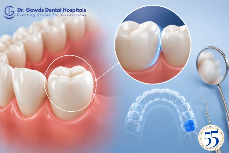
When a dentist describes a cavity, filling, tooth fracture, or gum problem, you may hear terms such as mesial, distal, buccal, lingual, or occlusal.
These terms describe specific surfaces or directions of a tooth. One of these is the distal tooth surface.
Although the term may sound technical, its meaning is relatively simple. The distal surface refers to the part of a tooth that is positioned away from the midline of the dental arch. The American Dental Association defines distal as the surface or position of a tooth most distant from the median line of the arch.
Understanding the distal surface can help patients make sense of dental charts, X-ray reports, treatment plans, and conversations with their dentist.
The distal area can also be clinically important because problems such as tooth decay, plaque accumulation, gum inflammation, fractures, and food trapping may involve surfaces between neighboring teeth.
At Dr Gowds Dental Hospitals, accurately identifying the affected tooth surface helps dental professionals assess the location and extent of a dental problem and plan appropriate treatment.
The distal tooth surface is the surface of a tooth that is positioned farther away from the midline of the dental arch.
Imagine an imaginary line running through the center of the dental arch between the two central incisors. As you move away from this midline along the dental arch, the surfaces positioned farther from it are described as distal.
For example:
The neighboring tooth varies depending on which tooth is being examined.
The easiest way to understand the distal surface is to compare it with the mesial surface.
The mesial surface is the side of a tooth positioned closer to the midline of the dental arch.
The distal surface is the side positioned farther away from the midline.
A simple way to remember the difference is:
Mesial = toward the middle
Distal = away from the middle
These terms allow dental professionals to describe the location of a dental problem more precisely.
Dentistry requires accurate descriptions of tooth anatomy.
Simply saying that a problem is on the “back side” or “side” of a tooth may be unclear because teeth have different positions and surfaces.
The term distal provides a consistent directional description that can be used across different teeth.
For example, a dentist may document:
These descriptions indicate where the problem is located.
The ADA also uses standardized tooth-surface terminology and abbreviations for dental documentation.
On front teeth, the distal surface is the side positioned farther away from the centerline of the dental arch.
For example, each central incisor has a mesial surface facing toward the dental midline and a distal surface facing away from it.
The same principle applies to the lateral incisors and canines.
The important point is that distal does not simply mean left or right. It describes the tooth’s position in relation to the midline.
On premolars and molars, the distal surface generally faces toward the back of the dental arch.
For example, the distal surface of a first molar generally faces toward the second molar.
The distal surface of a second molar faces toward the third molar when a wisdom tooth is present.
Because these areas are located between neighboring teeth, they may sometimes be difficult to clean thoroughly.
Neighboring teeth normally touch at contact areas.
The contact between a tooth and the tooth positioned farther back in the dental arch can be described in relation to its distal surface.
Healthy tooth contacts contribute to the stability and normal arrangement of the dental arch.
If the contact changes because of tooth decay, a restoration, tooth movement, or tooth loss, food may become trapped more easily.
Food trapping can make cleaning more difficult and may contribute to plaque accumulation.
The distal surface itself does not automatically cause tooth decay.
However, areas between neighboring teeth can be difficult to clean. Plaque can accumulate in these interdental areas, particularly when cleaning between the teeth is inadequate.
Over time, plaque bacteria can contribute to the development of dental caries.
The American Dental Association notes that interdental cleaning helps remove plaque and debris from areas between teeth and can help reduce the likelihood of tooth decay and gum disease.
A cavity involving the distal surface may also go unnoticed because it can be difficult to see directly.
Distal caries refers to tooth decay involving the distal surface of a tooth.
The decay may be limited to the distal surface or may extend to another surface.
For example, a dentist may describe decay as distal-occlusal, meaning that the affected area involves both the distal and occlusal surfaces of a posterior tooth.
The exact surfaces involved help the dentist understand the extent of the lesion and plan appropriate treatment.
A small cavity involving the distal surface may cause no noticeable symptoms.
As tooth decay progresses, symptoms may include:
More advanced decay can affect deeper structures of the tooth and may require more extensive treatment.
This is one reason regular dental examinations are useful even when you do not have tooth pain.
A cavity affecting the distal surface may develop between two teeth or in an area that is difficult to see directly.
You may therefore have no obvious visible hole or dark spot.
Depending on the situation, a dentist may use:
X-rays can be particularly useful for identifying decay between teeth that may not be visible during a routine visual examination.
Yes. Cleaning between teeth is an important part of maintaining oral health.
Brushing cleans many accessible tooth surfaces, but a toothbrush cannot effectively clean every area between adjacent teeth.
Interdental cleaning helps remove plaque and food debris from these hard-to-reach areas.
Depending on your individual dental needs, a dentist may recommend:
The most appropriate method depends on factors such as tooth spacing, gum health, dental restorations, and individual oral hygiene needs.
The areas around and between teeth are also important for gum health.
Plaque accumulation can contribute to gum inflammation.
Possible signs of gum problems include:
Some areas toward the back of the mouth may be difficult to access effectively with a toothbrush.
Regular interdental cleaning and professional dental care can help maintain healthy teeth and gums.
Treatment depends on the cause and severity of the problem.
A distal surface may be affected by:
A small cavity may require a filling.
A larger defect may require a more extensive restoration.
If the tooth has significant structural damage or deep infection, additional treatment may be necessary.
The word distal describes the location, not the treatment.
When a filling involves the distal surface, the dentist needs to restore the missing tooth structure while maintaining an appropriate relationship with the neighboring tooth.
The restoration should allow the area to be cleaned effectively and should preserve appropriate tooth anatomy and contact.
An improperly shaped restoration may contribute to:
The objective is therefore not simply to fill the cavity but to restore the tooth’s function and anatomy as appropriately as possible.
You may occasionally see abbreviations such as MOD in dental records.
MOD stands for:
An MOD restoration or lesion involves the mesial, occlusal, and distal surfaces of the tooth.
The ADA uses these standardized surface abbreviations in dental documentation.
Other combinations can also be used. For example, MO refers to mesial and occlusal surfaces, while DO refers to distal and occlusal surfaces.
These terms describe different aspects of tooth anatomy.
The surface positioned away from the midline of the dental arch.
The chewing surface of a posterior tooth, such as a premolar or molar.
A dental problem can involve one surface or multiple surfaces.
For example, a distal-occlusal cavity affects both the distal and occlusal surfaces of a posterior tooth.
No.
The appropriate treatment depends on what is affecting the tooth and how advanced the condition is.
For example, early noncavitated caries lesions may sometimes be managed with preventive or remineralization-focused approaches, depending on the patient’s individual risk and clinical findings. The ADA’s caries guidance distinguishes between noncavitated and cavitated lesions and considers factors such as tooth surface and dentition when determining management.
An established cavity may require restorative treatment.
A fractured distal surface may require a different type of restoration.
Therefore, distal describes where the problem is, not what treatment is automatically required.
The concept of distal surfaces also applies to primary teeth.
Children can develop cavities between baby teeth, and these areas may be difficult for parents to see.
Regular dental examinations can help identify problems at an earlier stage.
Primary teeth are important for chewing, speech, oral development, and maintaining space for permanent teeth. Therefore, dental problems in baby teeth should not automatically be ignored simply because those teeth will eventually be replaced.
A pediatric dentist or dental professional can determine whether preventive care, monitoring, restoration, or another treatment approach is appropriate.
The distal area of a second molar becomes particularly relevant when a wisdom tooth is present.
The area between the second and third molars may be difficult to clean effectively.
Partially erupted wisdom teeth can also create areas where plaque and food debris accumulate.
You should consider a dental examination if you experience:
A dentist can determine whether the wisdom tooth, the second molar, or another condition is responsible for the symptoms.
Dental treatment is often planned according to the specific tooth structures involved.
Knowing whether a problem is mesial, distal, occlusal, buccal, lingual, or another surface helps the dental team communicate the location accurately.
This can help determine:
Standardized dental terminology also supports consistent documentation and communication between dental professionals. The ADA’s CDT system is designed to support consistent documentation of dental treatment.
The distal tooth surface is the part of a tooth positioned away from the midline of the dental arch.
Although it may seem like a small anatomical detail, identifying the distal surface accurately is important in everyday dentistry. Dentists use this terminology when describing cavities, restorations, fractures, gum problems, and other dental conditions.
Distal surfaces can be particularly relevant when problems develop between neighboring teeth, where plaque and food debris may be more difficult to remove and early decay may not be immediately visible.
Understanding the difference between mesial and distal surfaces can also help patients better understand dental charts, X-ray reports, and treatment recommendations.
Good oral hygiene remains an important part of protecting these areas. Brushing twice daily with fluoride toothpaste, cleaning between the teeth, maintaining a tooth-friendly diet, and attending recommended dental examinations can help reduce the risk of dental problems.
If your dentist tells you that you have a problem on the distal surface of a tooth, remember that “distal” describes the location of the problem. The appropriate treatment depends on what is affecting the area and how advanced the condition is.
At Dr Gowds Dental Hospitals, detailed examination and individualized treatment planning can help identify and manage dental problems while preserving as much healthy tooth structure as possible.
Visit us at-
Dr gowds dental hospital Nanakramguda
Dr gowds dental hospital Gachibowli
Dr gowds dental hospital madhapur
The distal tooth surface is the part of a tooth positioned farther away from the midline of the dental arch.
Distal caries is tooth decay involving the distal surface of a tooth. The decay may also extend to another surface, such as the occlusal surface.
Cleaning between teeth helps remove plaque and food debris from areas that a toothbrush cannot effectively reach and can help reduce the risk of tooth decay and gum disease.
Distal-occlusal means that a dental problem or restoration involves both the distal surface and the occlusal chewing surface of a posterior tooth.
MOD stands for mesial, occlusal, and distal. It describes a lesion or restoration involving those three tooth surfaces.
No. Treatment depends on whether the area has early noncavitated caries, an established cavity, a fracture, or another dental problem.
Consider seeing a dentist if you experience tooth sensitivity, pain, food trapping, bleeding around a tooth, visible damage, or persistent discomfort. Regular dental examinations are also useful because some dental problems may not cause symptoms in their early stages.

 Gachibowli Branch